

KUWAIT: Dr Michael Masoomi, the lead researcher in the Department of Medical imaging - Adan Hospital, in association with research colleagues in Belgium, have managed to develop a 3D printing of a female human heart, for the first time in Kuwait and probably in the region.
"Three-dimensional printing has the potential for significant impact within healthcare, with its ability to create personalized healthcare and cost effective solutions," Dr Masoomi said. "The technology originated in engineering and the aeronautical industry that has begun to find applications in the world of medicine and has allowed the generation of accurate complex anatomical models."
The medical benefits of individualized 3D printed models include assisting clinical diagnosis, choosing the best operative strategy, predicting any intra-operative challenges in advance, education and training for junior surgeons, and generating customizable prostheses and implants to suit the individual patient.
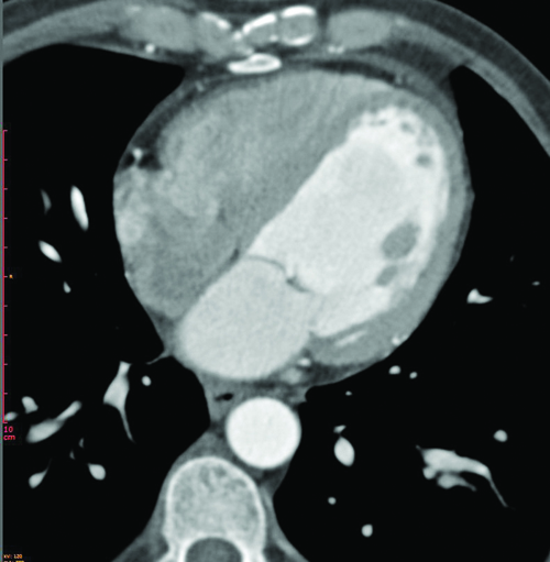
"The diagnosis and treatment of structural and congenital heart disease has traditionally relied on the analysis of images obtained by echocardiography, angiography, computed tomography (CT), and magnetic resonance imaging (MRI) and Nuclear Medicine imaging," Dr Masoomi said. In recent years, three-dimensional (3D) printed models have been incorporated into cardiology because of their potential usefulness in enhancing understanding of congenital heart disease, surgical planning, and simulation of structural percutaneous interventions.
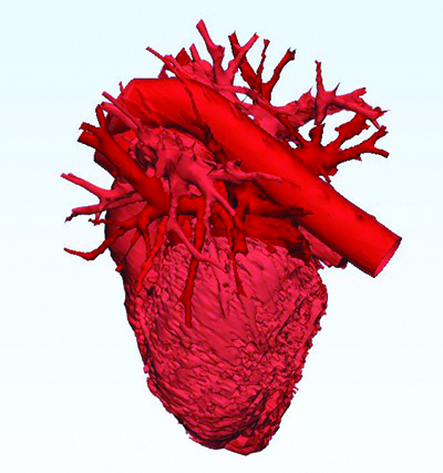
A 3D generated computer model of a female human heart. 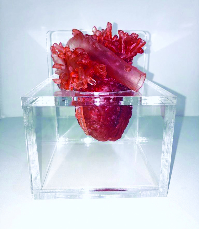
3D printing of a female human heart based on the generated computer model - Medical Imaging, Adan Hospital.
It also allows for the production of individualized cardiac stents to reduce the rate of in-stent re-stenosis. This application of 3D printing is especially relevant to the teaching of congenital heart disease because it can expose students to the full spectrum of malformations and the variability.
Moreover, the interconnectivity provided by the internet makes it easy to share digital files through online networks, allowing users to rapidly access and print 3D models of the disease of interest. Making 3D images available through online collections can provide hospitals and research centers with free access to a broad spectrum of heart conditions. Even more importantly, 3D models promise to transform teaching in ways that go beyond the lecture hall, and over the next few years are set to revolutionize medical training, especially in percutaneous interventions.
It has been reported there are current moves to make simulations a required element of cardiac catheterization training, part of a joint effort affecting the clinical practice guidelines of several international societies, including the Cardiovascular and Interventional Radiological Society of Europe, the Society for Cardiovascular Angiography and Interventions, the Society of Interventional Radiology, and the Radiological Society of North America.
Simulations involving 3D cardiac models allow trainees to practice with a diverse range of scenarios, repeat technical procedures, and learn from mistakes without putting patients at risk. Simulations can be used to train specialists before they carry out their first procedures on patients, for training reinforcement, and for instruction in new and more complex procedures.
Another application of 3D models is as a tool for explaining procedures to patients during medical consultations, a practice demonstrated to improve patient satisfaction and instill a sense of involvement with medical staff. Dr Latifah Al-Kandari, the Head of Medical Imaging Department, elaborated that there is no doubt that 3D printing is an immensely promising technology, set to have an enormous impact on medicine generally and in the treatment of congenital and structural heart diseases in particular. Widespread incorporation of 3D printing will be a major advantage in tackling new diagnostic and therapeutic challenges in the following years.